Topic 1 of 9

Research & data · Research dossier

Why we research delphinidin.
And how we measure.

Explore the research dossier ↓

At a glance

Starting point

Cells are exposed to oxidative stress in everyday life. This means an imbalance between reactive oxygen species (ROS) and the cell's regulatory systems. Delphinidin is a plant pigment from the group of anthocyanidins; here it comes from the maqui berry and is a chemically sensitive molecule: light, heat and oxygen make it break down quickly.

Research question

What role delphinidin plays in cell models is the subject of our research. We investigate this, among others, in kidney cells and in an intestinal cell line under defined, established experimental conditions. A second question concerns the complex itself: how its four delphinidin compounds can be detected analytically. Whether statements about humans can be derived from cell models is a separate, open question that is being worked on.

Methods

An insight into our work. Analytics: LC-MS of the four delphinidin compounds. Cell models: fluorescence microscopy in kidney cells, comet assay in an intestinal cell line, micronucleus test (microscopy, 400×). Pharmacokinetics: plasma levels in humans, with LC-MS at the core (internal study).

Limits

The figures show which investigations were carried out. They do not make a claim about an effect of CELL PREVAID® in humans. For the ongoing clinical studies, the protocols, design, research questions and results remain confidential for now. Publication is planned.

What makes PREVAID® distinctive

From the question to testing in humans.

Since 2008, a question from organ protection has become our own delphinidin complex: examined in the laboratory, patent pending, made in Germany.

1 · The origin

More than 40 years of anaesthesiological research into organ protection led to the question: can the sensitive plant pigment delphinidin be examined more closely at the cell? The cell model puts questions to the cell, analytics measures by LC-MS. The source: the maqui berry from Chile.

The question

Cells are under stress – in everyday life and especially during major surgery. From more than 40 years of Prof. Roewer's anaesthesiological research into how tissue can be preserved came the question whether the plant pigment delphinidin could be examined more closely at the cell.

Delphinidin

A plant pigment from the anthocyanidin group comes into view as a candidate; sensitive to light, heat and oxygen.

Cell model

Fluorescence microscopy, comet assay & micronucleus test: these methods answer questions about cells, within the limits of each method. Delphinidin remains the candidate we continue to pursue.

Analytics

LC-MS makes the identity and content of the delphinidin compounds measurable – the basis for optimising formulation and stability.

Maqui berry

Aristotelia chilensis from Chile, rich in anthocyanins – the source in which delphinidin is abundant.

2 · The solution

Our own process binds delphinidin to a ring-shaped sugar – the complex, patent pending. The ring protects the molecule from light, heat and oxygen. Analytics captures four delphinidin compounds individually, in every batch; the cell model now puts its questions to the finished formulation.

Complex

Our own process: delphinidin is bound to a ring-shaped sugar (alpha-cyclodextrin) and standardised. The ring protects the sensitive molecule from light, heat and oxygen and makes it stable and standardisable. Improved uptake has been demonstrated by our investigations. We are continuing to investigate the precise extent of this improvement in humans. Patent pending.

Analytics

The complex is characterised by LC-MS: four delphinidin compounds individually, every batch laboratory-tested.

Cell model

The complex returns to the cell model: the same questions put to the cell, now to the finished formulation.

3 · The test

Translation: what was measured in the laboratory is tested in humans. The product is developed and manufactured in Germany. In humans, an internal study measured the plasma levels over the day. Our clinical studies are currently under way, with details still confidential (publication planned).

Translation

Translation: the step from the laboratory to humans. What was measured in cells and in the analytics says nothing yet about how the complex behaves in the body after intake. So the first question is bioavailability in humans – measured, not assumed. Only on that basis can clinical studies be planned properly.

Product

Developed and manufactured in Germany, standardised and laboratory-tested.

Human

After analytical work and cell models: questions to explore in humans. Clinical investigations complement our analytical work and cell models with findings in humans. We are continuing to investigate the precise extent of the improvement in uptake.

Plasma levels

Pharmacokinetics in an internal proof-of-concept study: volunteers took the delphinidin complex; plasma levels were measured over the day according to a fixed protocol, analysed with LC-MS at the core and complemented by further analyses. The study is confidential. The results have yet to be published, but publication is planned.

Clinical studies

Our clinical studies are under way; questions, study design and results remain confidential for now.

From the first question to testing in humans: origin, solution, test – one research programme. Developed and manufactured in Germany.

A sample of our past work

The research dossier.

Analytics, cell models, plasma levels. A journey through our research. A diagram of the cell model comes first.

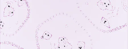
What we investigate at the cellular level.00 / 45 s
Schematic cell model

Cellular research

Cells are exposed to oxidative stress in everyday life.

45 s

A simplified illustration of complex biological relationships – not a statement about any effect of the product.

Understanding the illustration

The transition shows the same modelled cell geometry in schematic and fluorescent visual styles. Both are illustrations, not microscopy images or measured data. They do not establish any effects of CELL PREVAID®.
Explore the research question →

Diagram / Cell model · illustration

What we investigate at the cellular level.

A simplified illustration of complex biological relationships – not a statement about any effect of the product.

Context
The transition shows the same modelled cell geometry in schematic and fluorescent visual styles. Both are illustrations, not microscopy images or measured data. They do not establish any effects of CELL PREVAID®.
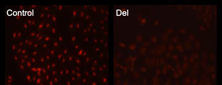
Four red fluorescence images labelled Control, Del, AMA and Del+AMA.

Fluorescence images from four of the six experimental groups of the original figure.

Cell model. Direct extrapolation to humans is not possible. This illustrates the method and does not present the full study protocol. No statement is made about an effect of CELL PREVAID®.

Fig. 1 · Fluorescence images from four of the six experimental groups of the original figure.

Insight / Fluorescence microscopy

Mitochondrial ROS formation in kidney cells

Fluorescence investigations of mitochondrial ROS formation in kidney cells. ROS stands for reactive oxygen species; a fluorescent dye makes their formation in the mitochondria visible under the microscope.

Research question
We investigate how mitochondrial ROS formation in kidney cells presents under a stressor (antimycin A) and what the experimental groups with delphinidin look like in the fluorescence image.
Method, limits and source
Method
Kidney cells. Control: control · Del: delphinidin · AMA: antimycin A. Group labels are retained from the original figure.
Limits and source
Limits
Cell model. Direct extrapolation to humans is not possible. This illustrates the method and does not present the full study protocol. No statement is made about an effect of CELL PREVAID®.
Source
Exemplary excerpts from internal research projects.
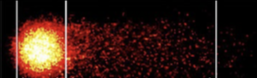
Supplied comet image with marked Head and Tail regions.

Comet image with marked head and tail.

Cell model. Direct extrapolation to humans is not possible. This illustrates the method and does not present the full study protocol. No statement is made about an effect of CELL PREVAID®.

Fig. 2 · Comet image with marked head and tail.

Insight / Comet assay

Comet assay: DNA strand breaks in an intestinal cell line

Investigation of H₂O₂-induced DNA strand breaks in an intestinal cell line. H₂O₂ (hydrogen peroxide) is used in the experiment to induce oxidative stress in a controlled way. In the comet assay, individual cell nuclei are embedded in a gel and exposed to an electric field. DNA fragments migrate out of the nucleus and form a tail – hence the name.

Research question
We investigate how H₂O₂-induced DNA strand breaks present in an intestinal cell line and what cells look like that were treated with delphinidin beforehand.
Method, limits and source
Method
Comet assay (single-cell gel electrophoresis) in an intestinal cell line; investigation of H₂O₂-induced DNA strand breaks. Head: cell nucleus · Tail: migrated DNA fragments.
Limits and source
Limits
Cell model. Direct extrapolation to humans is not possible. This illustrates the method and does not present the full study protocol. No statement is made about an effect of CELL PREVAID®.
Source
Exemplary excerpts from internal research projects.
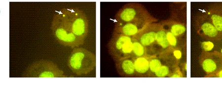
Three green-yellow microscopy panels with white arrows.

Three microscopy panels, 400×; arrows mark micronuclei.

Cell model. Direct extrapolation to humans is not possible. This illustrates the method and does not present the full study protocol. No statement is made about an effect of CELL PREVAID®.

Fig. 3 · Three microscopy panels, 400×; arrows mark micronuclei.

Insight / Micronucleus test

Micronucleus test: micronuclei under the microscope

Micronucleus test: microscopic examination of micronuclei at 400× magnification. Micronuclei are small additional nuclear bodies that can arise during cell division from chromosome fragments or whole chromosomes. The test records how frequently they occur in a cell population.

Research question
We investigate how frequently micronuclei occur after a challenge and how the frequency presents across the experimental groups.
Method, limits and source
Method
Micronucleus test; microscopic image at 400× magnification.
Limits and source
Limits
Cell model. Direct extrapolation to humans is not possible. This illustrates the method and does not present the full study protocol. No statement is made about an effect of CELL PREVAID®.
Source
Exemplary excerpts from internal research projects.

Insight / Analytics

Analytics: four delphinidin compounds in LC-MS

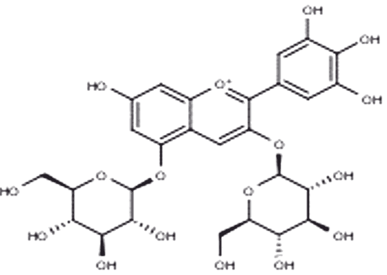
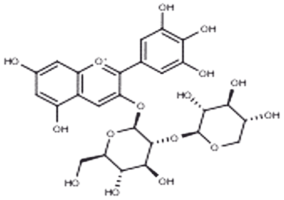
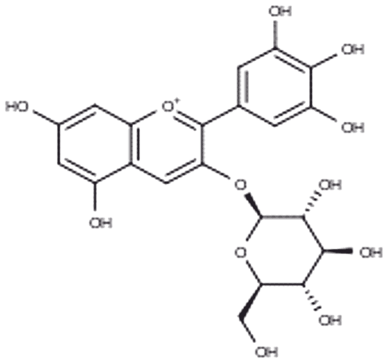
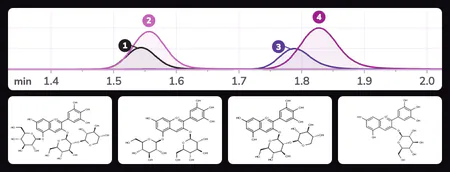
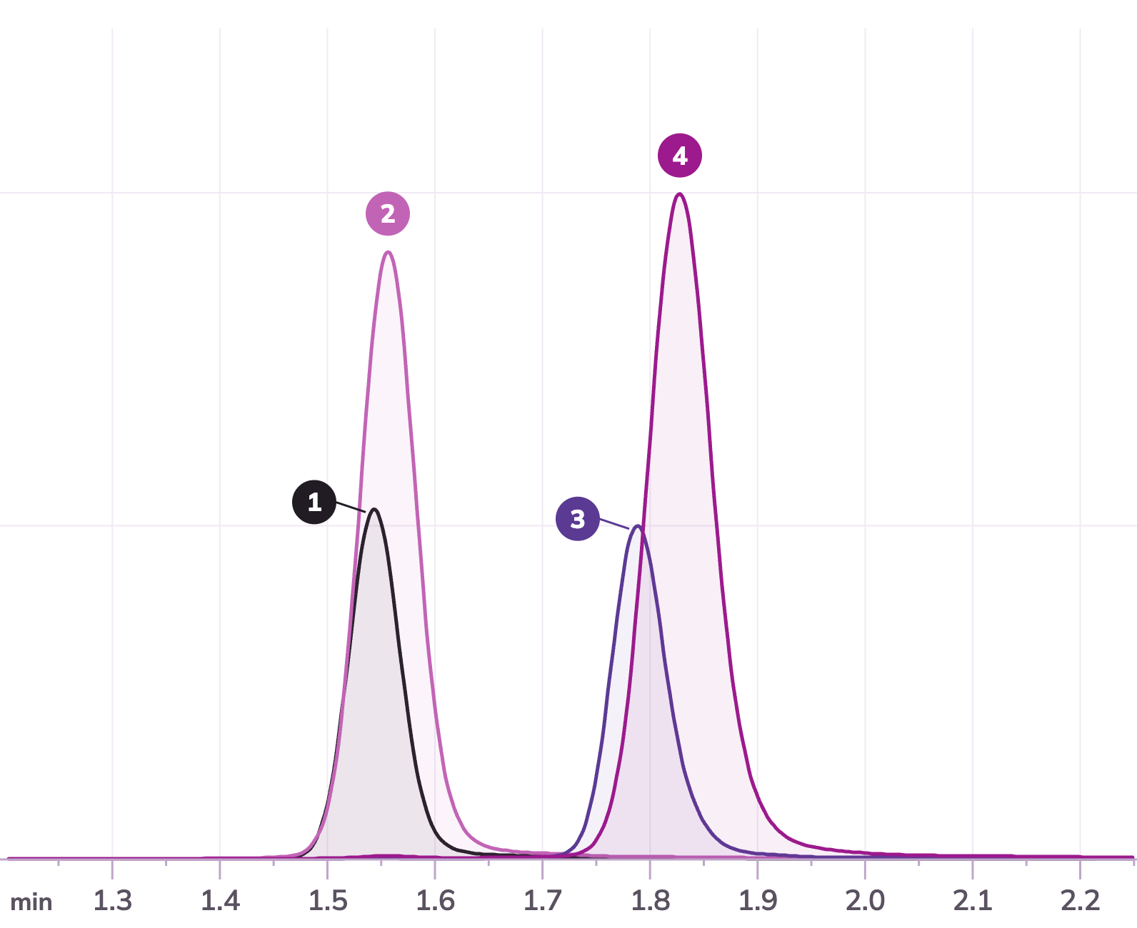
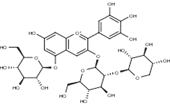
Analytical presentation of the compounds investigated using liquid chromatography coupled with mass spectrometry (LC-MS), a method for separating, identifying and quantifying chemical substances in complex mixtures. Liquid chromatography separates the components of a mixture so that each compound reaches the detector at its own time. The mass spectrometer then determines the mass of the molecules and so allows each curve to be assigned to a compound. Shown are four delphinidin compounds: delphinidin-3-glucoside, delphinidin-3,5-diglucoside, delphinidin-3-sambubioside and delphinidin-3-sambubioside-5-glucoside.

Research question
We investigate which delphinidin compounds the complex contains and how each can be detected and assigned individually.
Method, limits and source
Method
Liquid chromatography coupled with mass spectrometry (LC-MS); delphinidin complex, 25 µg/ml in 0.1 N HCl. Four compounds are assigned by retention time and mass transition.
Limits and source
Limits
Analytical characterisation of the complex. No statement about any effect of CELL PREVAID® in humans.
Source
Exemplary excerpts from internal research projects.
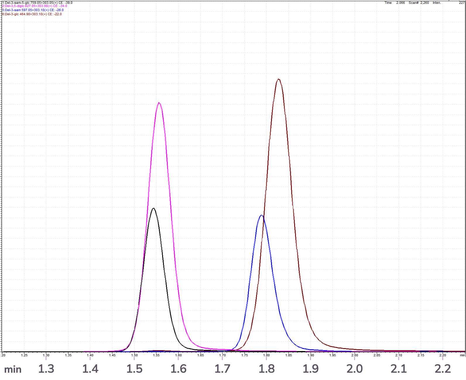
Chromatogram from the measuring software: four signal curves over time from 1.2 to 2.25 minutes, with the instrument legend at the top left.The same four curves redrawn and numbered: 1 and 2 peak at about 1.55 minutes, 3 and 4 at about 1.8 minutes.
Chromatogram of the delphinidin complex (25 µg/ml in 0.1 N HCl). The original image comes from the measuring software; for legibility we lightened the background, strengthened the lines and labelled the time axis in larger type. The explanation shows the same curves redrawn and numbered.

Each numbered curve stands for one of the compounds investigated.

In the mass spectrometer all four break down to the same delphinidin core (m/z 303); they differ only in their sugar moiety.

  1. 1

    Del-3-sam-5-glc

    Delphinidin 3-sambubioside-5-glucoside

    ≈ 1.54 min · m/z 759 → 303

    Structural formula: Delphinidin 3-sambubioside-5-glucoside
  2. 2

    Del-3,5-diglc

    Delphinidin 3,5-diglucoside

    ≈ 1.56 min · m/z 627 → 303

    Structural formula: Delphinidin 3,5-diglucoside
  3. 3

    Del-3-sam

    Delphinidin 3-sambubioside

    ≈ 1.79 min · m/z 597 → 303

    Structural formula: Delphinidin 3-sambubioside
  4. 4

    Del-3-glc

    Delphinidin 3-glucoside

    ≈ 1.83 min · m/z 465 → 303

    Structural formula: Delphinidin 3-glucoside

Pharmacokinetics · internal study

Plasma levels in humans

In an internal proof-of-concept study, volunteers took the delphinidin complex. Plasma levels were measured over the day according to a fixed protocol.

Research question
Whether, and over what time course, the delphinidin compounds of the complex can be detected in blood plasma after intake.
Method, limits and source
Method
Intake by volunteers, blood samples over the day according to a fixed protocol; at the core, determination of the delphinidin compounds in plasma by LC-MS, complemented by further analyses.
Limits and source
Limits
Internal study, measured values confidential; no statement about any effect of CELL PREVAID® in humans.
Source
Internal proof-of-concept study, not published; publication planned.

The figures give an insight into the scope of the investigations carried out. Original scientific data remain confidential.

Background for further reading

Two pages that deepen the dossier.

The literature behind it.

Publications in their respective research contexts – from reviews to cell and animal models to human studies. They show in which models delphinidin and related anthocyanins are investigated in the scientific literature. Their results apply to the respective model and cannot be transferred to CELL PREVAID®.

Show all 19 publicationsClose list ↑

10 papers

The compound: chemistry, stability, uptake

What anthocyanins and delphinidin are, how stable they are and how much of them is measured unchanged in the blood. These papers summarise what has been measured.

  1. Study type: Review

    Kumkum R, Aston-Mourney K, McNeill BA, Hernández D, Rivera LR.

    Bioavailability of Anthocyanins: Whole Foods versus Extracts.

    Nutrients. 2024;16(10):1403.

    PubMed ↗︎ (on PubMed, opens in a new tab)
  2. Study type: Review

    Gonçalves AC, Nunes AR, Falcão A, Alves G, Silva LR.

    Dietary Effects of Anthocyanins in Human Health: A Comprehensive Review.

    Pharmaceuticals (Basel). 2021;14(7):690.

    PubMed ↗︎ (on PubMed, opens in a new tab)
  3. Study type: Review

    Husain A, Chanana H, Khan SA, Dhanalekshmi UM, Ali M, Alghamdi AA, et al.

    Chemistry and Pharmacological Actions of Delphinidin, a Dietary Purple Pigment in Anthocyanidin and Anthocyanin Forms.

    Front Nutr. 2022;9:746881.

    PubMed ↗︎ (on PubMed, opens in a new tab)
  4. Study type: Review

    Avula B, Katragunta K, Osman AG, Ali Z, John Adams S, Chittiboyina AG, et al.

    Advances in the Chemistry, Analysis and Adulteration of Anthocyanin Rich-Berries and Fruits: 2000–2022.

    Molecules. 2023;28(2):560.

    PubMed ↗︎ (on PubMed, opens in a new tab)
  5. Study type: Review

    Noman AM, Sultan MT, Maaz M, Mazhar A, Tariq N, Imran M, et al.

    Nutraceutical Potential of Anthocyanins: A Comprehensive Treatise.

    Food Sci Nutr. 2025;13(5):e70164.

    PubMed ↗︎ (on PubMed, opens in a new tab)
  6. Study type: Review

    Valdez JC, Bolling BW.

    Anthocyanins and intestinal barrier function: a review.

    J Food Bioact. 2019;5:18-30.

  7. Study type: Review

    Zhang L, Wang Y, Cao Y, Wang F, Li F.

    Review: Enhancing the Bioavailability and Stability of Anthocyanins for the Prevention and Treatment of Central Nervous System-Related Diseases.

    Foods. 2025;14(14):2420.

    PubMed ↗︎ (on PubMed, opens in a new tab)
  8. Study type: Animal model + in vitro

    Hidalgo J, Teuber S, Morera FJ, Ojeda C, Flores CA, Hidalgo MA, et al.

    Delphinidin Reduces Glucose Uptake in Mice Jejunal Tissue and Human Intestinal Cells Lines through FFA1/GPR40.

    Int J Mol Sci. 2017;18(4):750.

    PubMed ↗︎ (on PubMed, opens in a new tab)
  9. Study type: Review

    Oliveira H, Fernandes A, Brás NF, Mateus N, de Freitas V, Fernandes I.

    Anthocyanins as Antidiabetic Agents—In Vitro and In Silico Approaches of Preventive and Therapeutic Effects.

    Molecules. 2020;25(17):3813.

    PubMed ↗︎ (on PubMed, opens in a new tab)
  10. Study type: Review

    Li S, Wu B, Fu W, Reddivari L.

    The Anti-inflammatory Effects of Dietary Anthocyanins against Ulcerative Colitis.

    Int J Mol Sci. 2019;20(10):2588.

    PubMed ↗︎ (on PubMed, opens in a new tab)

6 papers

Research in cell and animal models

Laboratory work in which research groups study anthocyanins in cells and animal models. The titles name the respective model; results from models are not a statement about humans.

  1. Study type: Review

    Sadowska-Bartosz I, Bartosz G.

    Antioxidant Activity of Anthocyanins and Anthocyanidins: A Critical Review.

    Int J Mol Sci. 2024;25(22):12001.

    PubMed ↗︎ (on PubMed, opens in a new tab)
  2. Study type: Review

    Ma Z, Du B, Li J, Yang Y, Zhu F.

    An Insight into Anti-Inflammatory Activities and Inflammation Related Diseases of Anthocyanins: A Review of Both In Vivo and In Vitro Investigations.

    Int J Mol Sci. 2021;22(20):11076.

    PubMed ↗︎ (on PubMed, opens in a new tab)
  3. Study type: In vitro

    Song SE, Jo HJ, Kim YW, Cho YJ, Kim JR, Park SY.

    Delphinidin prevents high glucose-induced cell proliferation and collagen synthesis by inhibition of NOX-1 and mitochondrial superoxide in mesangial cells.

    J Pharmacol Sci. 2016;130(4):235-243.

    PubMed ↗︎ (on PubMed, opens in a new tab)
  4. Study type: Animal model + in vitro

    Chamcheu JC, Adhami VM, Esnault S, Sechi M, Siddiqui IA, Satyshur KA, et al.

    Dual Inhibition of PI3K/Akt and mTOR by the Dietary Antioxidant, Delphinidin, Ameliorates Psoriatic Features In Vitro and in an Imiquimod-Induced Psoriasis-Like Disease in Mice.

    Antioxid Redox Signal. 2017;26(2):49-69.

    PubMed ↗︎ (on PubMed, opens in a new tab)
  5. Study type: Animal model + in vitro

    Liu Y, Hong T, Lv M, Guo X, Zhang P, Yan A, et al.

    Delphinidin attenuates cognitive deficits and pathology of Alzheimer’s disease by preventing microglial senescence via AMPK/SIRT1 pathway.

    Alzheimers Res Ther. 2025;17(1):138.

  6. Study type: Review

    Jeyaraj EJ, Vidana Gamage GC, Cintrat JC, Choo WS.

    Acylated and non-acylated anthocyanins as antibacterial and antibiofilm agents.

    Discov Food. 2023;3(1):21.

3 papers

Background: cellular stress and mitochondria

Reviews and one observational study in humans on the starting point: oxidative stress and the role of mitochondria. They do not investigate delphinidin or anthocyanins.

  1. Study type: Review

    Caturano A, D’Angelo M, Mormone A, Russo V, Mollica MP, Salvatore T, et al.

    Oxidative Stress in Type 2 Diabetes: Impacts from Pathogenesis to Lifestyle Modifications.

    Curr Issues Mol Biol. 2023;45(8):6651-6666.

    PubMed ↗︎ (on PubMed, opens in a new tab)
  2. Study type: Review

    Iacobini C, Vitale M, Pesce C, Pugliese G, Menini S.

    Diabetic Complications and Oxidative Stress: A 20-Year Voyage Back in Time and Back to the Future.

    Antioxidants (Basel). 2021;10(5):727.

    PubMed ↗︎ (on PubMed, opens in a new tab)
  3. Study type: Human study

    Zampino M, Brennan NA, Kuo PL, Spencer RG, Fishbein KW, Simonsick EM, et al.

    Poor mitochondrial health and systemic inflammation? Test of a classic hypothesis in the Baltimore Longitudinal Study of Aging.

    Geroscience. 2020;42(4):1175-1182.

    PubMed ↗︎ (on PubMed, opens in a new tab)

Measure, don't claim.

Our research questions. Our methods. Our limits.

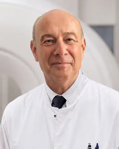
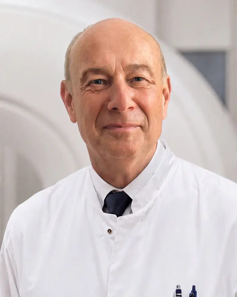
Prof. Norbert Roewer

Prof. Dr. med. Dr. h.c. Norbert Roewer

ROEWER MEDICAL CARE

The research behind PREVAID® connects questions about the natural compound and its formulation with scientific investigations: analytics on the complex, cell models in the laboratory, clinical studies in humans. The methods come from more than 40 years of anaesthesiological research by Prof. Roewer on organ protection, that is, the question of how tissue can be preserved during major surgery. The work is carried out at the Fraunhofer Translational Center for Regenerative Therapies in Würzburg and in university laboratories in Bochum, Munich, Essen and Würzburg.

“For us, innovation means wanting to respond to problems, to states of disease, with new approaches. In medicine, solutions and ideas need scientific craft as their foundation if they are to achieve real progress with foresight and courage. That is our credo.”

Prof. Dr. Norbert Roewer

Study status · ongoing

Clinical studies

After analytical work and cell models: questions to explore in humans.

Our clinical studies are ongoing. Details of the research questions, study designs and results remain confidential for now. Cell models and analytics answer laboratory questions. Clinical investigations complement our analytical work and cell models with findings in humans. We are continuing to investigate the precise extent of the improvement in uptake.